Table of Contents
Ultrasound imaging is a fundamental tool in medical diagnostics, offering a non-invasive glimpse into the human body to aid in the evaluation and diagnosis of various conditions. Among the various types of ultrasound, the transvaginal ultrasound (TVS) stands out for its unique application in women’s health. This procedure involves using sound waves to create images of the female reproductive organs, including the uterus, fallopian tubes, ovaries, cervix, and the pelvic area. Unlike traditional abdominal ultrasounds, transvaginal ultrasound provides a closer, more detailed view, making it an invaluable diagnostic tool for specific conditions.
The purpose of vaginal ultrasound is multifaceted. It is often recommended for investigating unexplained pelvic pain, abnormal bleeding, fibroids, cysts, and other gynecological issues. It plays a crucial role in early pregnancy assessments, fertility treatments, and the detection of ectopic pregnancies. Its ability to provide detailed images is particularly beneficial in diagnosing and monitoring conditions that cannot be as easily assessed through an abdominal ultrasound.
The safety of ultrasound technology is well-documented, offering a reassuring option for those undergoing diagnostic procedures. Sound waves, rather than radiation, are used to create images, positioning ultrasound as a preferable choice for many patients and healthcare providers alike.
Transvaginal ultrasounds are a routine part of gynecological and prenatal care, reflecting their significant role in maintaining women’s health. A study published in the Journal of Ultrasound in Medicine highlights the procedure’s effectiveness in early pregnancy evaluation, demonstrating its critical role in medical diagnostics. Similarly, research featured in Obstetrics & Gynecology outlines how transvaginal ultrasound is instrumental in identifying ovarian tumors, showcasing the procedure’s broad applicability.
For individuals preparing for or considering a transvaginal ultrasound, understanding the procedure, its benefits, and what to expect can alleviate concerns and facilitate a smoother diagnostic experience. This guide aims to provide comprehensive insights into transvaginal ultrasound, ensuring that patients are well-informed and comfortable with their upcoming procedure.
What is Transvaginal Ultrasound?
Transvaginal ultrasound also know as internal ultrasound is a type of medical imaging technique that uses sound waves to create pictures of the inside of the pelvic area, primarily focusing on a woman’s reproductive system, including the uterus, fallopian tubes, ovaries, cervix, and vaginal area. Unlike traditional ultrasound methods that are performed by moving a device across the surface of the abdomen, transvaginal ultrasound involves the use of a specially designed probe that is gently inserted into the vagina. This method provides a closer view of the pelvic organs and structures, offering clearer and more detailed images.
One of the main advantages of transvaginal ultrasound is its ability to produce high-quality images that help healthcare providers diagnose and monitor conditions that affect women’s reproductive health. For example, it plays a critical role in identifying abnormalities such as ovarian cysts, uterine fibroids, and ectopic pregnancies. It is also extensively used in fertility treatments and early pregnancy assessments, providing valuable information about the health and development of the embryo or fetus.
The procedure is generally considered safe, with minimal risks. Studies have consistently shown that the use of ultrasound in medical imaging does not expose patients to radiation, making it a preferred diagnostic tool for pregnant women and those undergoing fertility treatments. A study published in the Journal of Ultrasound in Medicine highlighted the effectiveness of transvaginal ultrasound in early pregnancy diagnosis and its role in the accurate detection of fetal anomalies and complications.
Preparing for a transvaginal ultrasound usually requires minimal preparation. Patients are often advised to have a full bladder, which can enhance the quality of the images, although this is not always necessary, especially for transvaginal scans. The procedure is quick, typically taking about 30 minutes, and is performed in an outpatient setting. Patients might experience mild discomfort during the insertion of the probe, but severe pain is rare. Communicating any concerns or discomfort with the healthcare provider can help make the experience as comfortable as possible.
Transvaginal ultrasound has become a standard tool in gynecological care, praised for its accuracy, safety, and non-invasiveness. Its ability to provide essential insights into the health and function of the pelvic organs makes it an invaluable diagnostic tool. As medical technology advances, the applications and capabilities of transvaginal ultrasound continue to expand, offering hope and answers to many women experiencing reproductive health issues.
Why is Transvaginal Ultrasound Done?
Transvaginal ultrasound (TVU or TVS) serves as a crucial diagnostic tool across various aspects of women’s health, offering insights into conditions that are often not detectable through external examination methods. Its primary purposes span from the assessment of pelvic pain, unexplained bleeding, and reproductive health issues to guiding fertility treatments. Here are some specific reasons why transvaginal ultrasound is performed:
Diagnosis of Gynecological Conditions: endovaginal ultrasound plays a pivotal role in diagnosing conditions such as ovarian cysts, uterine fibroids, and endometrial abnormalities. It provides detailed images of the uterus, ovaries, and other pelvic organs, which helps in identifying the cause of pelvic pain or abnormal bleeding. A study published in Minerva medica highlighted the sensitivity and specificity of TVU in detecting myometrial infiltration and cervical invasion in endometrial cancer patients, comparing favorably to MRI but at lower costs and with greater patient tolerability (Capozzi et al., 2020).
Fertility Assessment and Pregnancy Monitoring: For women undergoing fertility evaluations or treatments, the TVS sonogram is indispensable. It allows for the observation of the ovaries to monitor follicle development during ovulation induction and assists in procedures such as egg retrieval for in vitro fertilization. Furthermore, TVU is fundamental in early pregnancy for confirming viability, identifying ectopic pregnancies, and evaluating the cause of any first-trimester bleeding. The accuracy of TVU in diagnosing deep infiltrating endometriosis before surgery, which can affect fertility, was also emphasized in a study published in the Journal of Ultrasound in Medicine (Deslandes et al., 2020).
Screening for Ovarian Cancer: In an asymptomatic screening population, the role of transvaginal ultrasonography has been systematically reviewed to determine its effectiveness in reducing mortality from ovarian cancer. Although the current studies do not show a benefit in screening with TVU annually, they highlight the need for further research to improve screening protocols and technologies (Buhling et al., 2017).
Evaluation of Postmenopausal Bleeding: TVU is a first-line investigation for women experiencing postmenopausal bleeding (PMB), allowing for the assessment of the endometrial thickness and identifying potential malignancies. Saline contrast sonohysterography, an advanced application of TVU, has shown improved diagnostic accuracy for delineating intracavitary structures in women with PMB (Tahmasebi et al., 2020).
Assessment of Placenta and Fetal Health in Pregnancy: TVU is employed to diagnose placenta previa and assess placental location accurately. It helps in planning the management based on placental localization, which is crucial for reducing risks associated with placenta previa during delivery (Reed et al., 2008).
Through these applications, transvaginal ultrasound has established itself as a versatile, non-invasive diagnostic tool that significantly contributes to the comprehensive care of women’s health issues. Its ability to provide detailed images of the pelvic organs makes it an invaluable asset in the diagnosis, treatment planning, and monitoring of various gynecological and obstetric conditions.
What does a Transvaginal ultrasound show?
A transvaginal ultrasound provides detailed images of the female pelvic organs and structures, including the uterus, cervix, ovaries, fallopian tubes, and the bladder. By using high-frequency sound waves emitted from a probe inserted into the vagina, this type of ultrasound allows healthcare providers to examine these organs in detail.
Preparing for Your Transvaginal Sonography
When you are scheduled for a transvaginal ultrasound, understanding how to prepare can make the experience smoother and more comfortable. This non-invasive diagnostic tool is widely used by healthcare professionals to obtain clear images of the female reproductive system, including the uterus, ovaries, and cervix. Preparing for the procedure involves a few simple steps that ensure the most accurate results.
Before the Ultrasound
Unlike some medical tests, transvaginal ultrasound preparation is minimal. You may be advised to drink water before your appointment to fill your bladder, which helps to push the uterus into a position that is easier to scan, providing clearer images. The specific amount of water recommended can vary, but generally, patients are asked to drink about two to three glasses of water an hour before the exam. It’s important not to urinate after drinking the water until after your examination, if possible.
Wearing comfortable, loose-fitting clothing to your appointment can make it easier for you to undress from the waist down, which is necessary for the procedure. Some clinics may provide you with a gown for added comfort and privacy during the ultrasound.
What to Expect
Upon arrival, you will likely be asked to lie down on a medical exam table, with your knees bent and feet placed in stirrups. A protective cover is placed over the ultrasound probe before it is gently inserted into the vagina. The probe is slender and should not cause significant discomfort. If you feel anxious about the procedure, communicating with your sonographer can help. They can explain each step in real-time, which might ease any discomfort or anxiety.
Special Considerations
For most people, there are no specific dietary restrictions or preparation requirements beyond what has already been mentioned. However, if you have a history of pelvic pain or concerns about discomfort during the procedure, discussing these with your healthcare provider beforehand can be beneficial. They may provide additional guidance or adjustments to make sure you are as comfortable as possible.
Research and clinical guidelines suggest that transvaginal ultrasound is a safe and effective method for examining the pelvic region. A study published in the Journal of Clinical Ultrasound confirms its utility in accurately diagnosing conditions that affect women’s reproductive health. This reassurance, backed by scientific evidence, underscores the procedure’s role in preventive health care and diagnosis.
Remember, the goal of a transvaginal ultrasound is to gain insights into your health in a way that is minimally invasive. Preparing yourself by following these simple steps can contribute to a more positive and informative experience. If you have any questions or concerns before your appointment, do not hesitate to contact your healthcare provider. They are there to ensure your comfort and care throughout the process.
During the Procedure
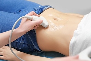
When undergoing a transvaginal ultrasound, the process is straightforward and designed with your comfort in mind. Before the procedure begins, you will be asked to empty your bladder. You will then be guided to a private room where you can change into a gown, ensuring your privacy and comfort throughout the examination. The procedure itself is performed with you lying on an examination table, with your knees bent and feet placed in stirrups, similar to the position during a gynecological exam.
A transvaginal ultrasound uses a small, wand-like device called a transducer, which is covered with a protective sheath and lubricated. The sonographer, a trained ultrasound technician, will gently insert the transducer into the vagina. Its size and shape are designed to minimize discomfort, and most individuals report feeling only slight pressure during insertion.
The transducer emits sound waves that bounce off the pelvic organs, creating images that are displayed on a monitor. The sonographer moves the transducer within the vaginal canal to obtain images from different angles, ensuring a comprehensive view of the uterus, ovaries, and other pelvic structures. This movement may cause varying sensations of pressure, but it is typically not painful.
Throughout the procedure, the sonographer may explain what is being done and can answer questions about the images being viewed. This can be reassuring and make the experience more informative. The procedure usually lasts between 15 to 30 minutes, depending on the specific reason for the examination.
Studies have shown that transvaginal ultrasound is a critical tool for assessing conditions within the female pelvis, offering detailed images that are crucial for accurate diagnosis and treatment planning. For instance, a study published in the American Journal of Obstetrics and Gynecology highlighted its effectiveness in evaluating ovarian masses, providing valuable information that guides clinical decisions.
After the examination, the sonographer will remove the transducer, and you will be given time to change back into your clothes. There may be a brief wait while the images are reviewed, and in some cases, a radiologist or your healthcare provider may discuss preliminary findings with you directly after the procedure.
Remember, the goal of a transvaginal ultrasound is to gain the clearest picture possible of your pelvic health. While it may feel a bit unusual, the procedure is a routine part of women’s healthcare, designed to be as comfortable and informative as possible.
Understanding Your Results
After undergoing a transvaginal ultrasound, the next step is to understand the results. The radiologist will review the images captured during the scan to assess for any abnormalities or conditions that could be affecting your reproductive health. This could include checking the health of your ovaries, uterus, fallopian tubes, and the surrounding pelvic area. The clarity and detail provided by transvaginal ultrasound allow for a more accurate diagnosis compared to other types of ultrasound imaging.
Results from transvaginal ultrasounds are typically ready within a few days, depending on the facility. Your doctor will schedule a follow-up appointment to discuss the findings with you. During this meeting, they will explain what the images reveal about your pelvic organs and if there are any signs of conditions such as cysts, fibroids, or other abnormalities. If the scan was part of fertility treatment or pregnancy monitoring, your doctor would discuss the health of the embryo or fetus and any observations relevant to your pregnancy.
Sometimes, the results might indicate the need for further testing or follow-up scans to monitor a condition or to get a clearer understanding of what was seen. This is common and should not be a cause for immediate concern. It’s just a step towards ensuring you receive the most accurate diagnosis and appropriate care.
A study published in the American Journal of Obstetrics and Gynecology highlighted the importance of transvaginal ultrasounds in early pregnancy for accurately dating the pregnancy and assessing fetal health. Another research article in the Journal of Clinical Ultrasound discussed how transvaginal ultrasound is crucial in diagnosing and managing ectopic pregnancies, demonstrating the procedure’s vital role in women’s health.
Your healthcare provider will use the results of your transvaginal ultrasound, along with other information from your medical history and possibly further tests, to create a comprehensive view of your reproductive health. This might involve recommending treatment options if any issues are identified or simply providing reassurance about your condition.
It’s important to ask questions during your follow-up appointment if anything about your results is unclear. Understanding the findings from your transvaginal ultrasound can help you make informed decisions about your health and treatment options moving forward.
Safety and Risks
Transvaginal ultrasound is a widely used diagnostic tool that plays a significant role in women’s health care. Its safety profile is well-established, with decades of clinical use underscoring its value. Unlike X-rays, transvaginal ultrasound does not use ionizing radiation. Instead, it employs sound waves to create images of the internal organs. This method is considered safe for all patients, including pregnant women, as supported by extensive research and guidelines from health organizations worldwide.
Despite its high safety standards, it is normal for patients to have concerns about potential risks or discomforts associated with the procedure. Most patients experience minimal to no discomfort during a transvaginal ultrasound. The sensation associated with the insertion of the ultrasound probe can vary among individuals, but it is generally described as a mild pressure rather than pain.
Occasionally, some patients might experience slight discomfort if the probe is manipulated to gain better views of certain areas. However, sonographers are skilled professionals trained to perform the procedure with utmost care, ensuring patient comfort while obtaining the necessary diagnostic images.
Concerns about privacy and modesty are also addressed with sensitivity. The procedure is conducted in a private room, and patients are draped to maintain their dignity throughout the examination. Patients have the right to ask for a female sonographer if that makes them more comfortable.
Myths and misconceptions can also contribute to patient apprehension. One common myth is that the procedure is excessively invasive and painful, which is not supported by the experiences of the majority of patients who undergo transvaginal ultrasounds. Education and clear communication from healthcare providers can significantly alleviate these concerns, providing patients with accurate information about what to expect.
A study published in the Journal of Ultrasound in Medicine highlighted the low incidence of adverse effects associated with transvaginal ultrasounds, reinforcing the procedure’s safety. The study emphasized the importance of patient education in reducing anxiety and increasing comfort levels during the ultrasound.
Patients are encouraged to discuss any concerns or questions they may have with their healthcare provider before the procedure. This dialogue ensures that patients are fully informed and comfortable with the process, contributing to a positive and reassuring experience.
The decision to undergo a transvaginal ultrasound is a collaborative one between a patient and their healthcare provider. It is based on the potential benefits of obtaining valuable diagnostic information that can guide treatment and health decisions. As with any medical procedure, individual considerations and health conditions will influence this decision, but the overarching consensus from the medical community supports the safety and efficacy of transvaginal ultrasound in contributing to comprehensive women’s healthcare.
Alternative imaging techniques to transvaginal ultrasound
When considering alternative imaging techniques to transvaginal ultrasound for evaluating the pelvis and reproductive organs, several options are available, each with its specific applications and advantages. Here’s an overview of alternative imaging modalities:
Transabdominal Ultrasound: Unlike the transvaginal approach, the transabdominal ultrasound is performed by moving a transducer across the abdomen. This method is less invasive and can provide a broader view of the abdominal cavity and pelvic area, though it may not offer the same level of detail for certain conditions.
Magnetic Resonance Imaging (MRI): MRI offers high-resolution images of the pelvic organs and structures without using ionizing radiation. It’s particularly useful for assessing complex gynecological conditions, endometriosis, uterine anomalies, and pelvic masses. MRI can provide detailed information on soft tissue contrast, making it an excellent tool for planning surgical interventions or further evaluating abnormalities detected by ultrasound.
Computed Tomography (CT) Scan: While not commonly used for pelvic examinations due to its lower sensitivity in distinguishing soft tissues compared to MRI and the concern of radiation exposure, CT scans can be helpful in evaluating the pelvis for certain conditions. It’s particularly useful for assessing pelvic fractures, complex appendicitis, and in oncological assessments where metastasis to pelvic bones or lymph node involvement is suspected.
Hysterosalpingography (HSG): This is a specific type of X-ray that looks at the inside of the uterus and fallopian tubes and the area around them. It often is used to investigate infertility by looking for blockages or other problems in the fallopian tubes.
Sonohysterography: Also known as saline infusion sonography, this technique involves the introduction of saline into the uterus via a catheter during ultrasound imaging. It enhances the visualization of the uterine cavity and endometrial lining, aiding in the diagnosis of uterine anomalies, polyps, and fibroids.
3D Ultrasound: Building on traditional ultrasound technology, 3D ultrasounds can provide detailed images of the uterus and ovaries in three dimensions. This can be particularly useful for assessing the shape and structure of reproductive organs, though it’s more commonly used in obstetrics.
Each of these imaging techniques has its own set of indications, advantages, and limitations. The choice of imaging modality depends on the specific clinical scenario, the area of interest, patient factors (such as pregnancy status or contraindications to certain types of imaging), and the information needed to guide diagnosis and treatment. Your healthcare provider will consider these factors when recommending the most appropriate imaging technique for your individual situation.
Frequently Asked Questions about Transvaginal Ultrasound
Transvaginal ultrasound is a procedure that many people have questions about, especially when facing it for the first time. This section aims to address some of the most common inquiries with straightforward answers.
Can transvaginal ultrasound detect all types of pelvic conditions?
While transvaginal ultrasound is a highly effective diagnostic tool for assessing many pelvic conditions, it’s not all-encompassing. Its ability to provide detailed images of the uterus, ovaries, and other pelvic structures makes it invaluable for diagnosing conditions like ovarian cysts, fibroids, and early pregnancy. However, certain conditions may require additional tests for a comprehensive evaluation.
Is the procedure painful?
Most individuals experience little to no discomfort during a transvaginal ultrasound. The probe used in the procedure is designed to be as slim and comfortable as possible. Some may feel a slight pressure as the probe is inserted, but this is generally not painful. Communicating any discomfort to the sonographer can help adjust the procedure for your comfort.
How should I prepare for my appointment?
Preparation for a transvaginal ultrasound is minimal. You may be advised to empty your bladder before the procedure, but specific instructions can vary based on the reason for the exam. Always follow the guidance provided by your healthcare provider or the imaging center.
What if the ultrasound finds something?
Discovering unexpected findings during an ultrasound can be concerning, but it’s important to remember that ultrasounds are a first step in diagnostic processes. If something is found, your healthcare provider will discuss the findings with you and recommend next steps, which may include further testing for a definitive diagnosis.
How long does the procedure take?
A typical transvaginal ultrasound usually takes between 5 to 10 minutes. The duration can vary depending on the complexity of the examination and the specific information being sought by the healthcare provider.
Will I be able to see the images during the procedure?
Many sonographers will explain the images during the procedure, offering you a chance to see the screen. However, interpreting these images requires specialized training, so your healthcare provider will provide a detailed explanation of the findings during a follow-up appointment.
Can I bring someone with me?
Policies on having someone accompany you during the procedure vary by facility. It’s best to check with the location where your ultrasound is scheduled. While privacy is a priority during the exam, understanding and accommodating personal comfort is also important.
Can you do a vaginal ultrasound if you are a virgin?
As the probe needs to be inserted in the vagina, the examination is contraindicated If you are a virgin (Virgo intacta; hymen intact). Other types of ultrasound imaging such as transabdominal ultrasounds (through the abdomen) or a transrectal (through the rectum) ultrasound might be a viable alternative. (You can find more information at https://clinicaltrials.gov/ct2/show/NCT04858919 and https://www.ncbi.nlm.nih.gov/pmc/articles/PMC4719089/ )
Does a pelvic ultrasound hurt a virgin?
Transvaginal ultrasound is not indicated if you are a virgin in the UK as it can be painful. Please note that there is a lot of outdated information online about transvaginal ultrasounds and virginity. We follow NHS guidelines, and we are not writing an article just from the information we found online.
What is the risk of a transvaginal ultrasound?
There are no known risks from the sound waves used in an ultrasound scan. Unlike some other scans, such as CT scans, ultrasound scans don’t involve exposure to radiation External and internal ultrasound scans don’t have any side effects and are generally painless, although you may experience some discomfort as the probe is pressed over your skin or inserted into your body.
Read more about what is an ultrasound scan.
If you’re having an internal scan and are allergic to latex, it’s important to let the sonographer or doctor carrying out the scan know this so that they can use a latex-free probe cover.
Performing transvaginal ultrasounds on pregnant women is also safe, for both mother and fetus. This is because no radiation is used in this imaging technique.
When an internal ultrasound can detect pregnancy?
The best time to confirm pregnancy with an internal scan is from 6 weeks.
Why can’t my ovaries be seen on a transvaginal ultrasound?
It is very common for the bowel to obscure the ovaries.
Is an internal vaginal ultrasound necessary during a routine pregnancy ultrasound?
It is only necessary if the abdominal ultrasound is inconclusive.
Can I have an internal ultrasound if I am bleeding?
It is absolutely fine to have an ultrasound while you are bleeding, as far as you are comfortable with that.
For those with additional questions or concerns about undergoing a transvaginal ultrasound, do not hesitate to reach out to your healthcare provider. They can provide personalized information and reassurance based on your medical history and the specific reason for the ultrasound. This dialogue ensures that you’re fully informed and comfortable with the process, reflecting the commitment to patient care and understanding.
Medically Reviewed by Tareq Ismail Pg(Dip), BSc (Hons)